Archive for the 'Arthropods' Category

It’s a trap!

Yesterday, I was poking around a small bush of white flowers looking for insects to photograph. I noticed this butterfly hanging from the bottom of a flower, rather than sitting on top:

What are you doing under there?

I panned around to underneath the flower and found out why:

Unlucky butterfly.

The butterfly had been snared by an ambush bug (Phymatinae), which is a subgroup of assassin bugs (Reduviidae). I think the above animal is a nymph belonging to the genus Phymata. These bugs hang out underneath flower petals until unweary pollinators visit. They then lunge out and snare their prey with their enlarged raptorial appendages, piercing the exoskeleton with a syringe like rostrum.

Here is an adult of the same, or a similar, species. About one of every three flowers in this bush had an ambush bug laying in wait below.

Phymata sp

'Come a bit closer my pretties.'

If any insect-gurus can identify this exact species, it would be much appreciated.

The Fastest Claw in the West

I’m surprised that I haven’t gotten around to posting this yet. Here is one of my favorite mantis shrimp videos of all time.

This segment was a bit of humor produced for ‘The Fastest Claw in the West,’ a documentary from 1985 about mantis shrimp. It is narrated by blog-hero David Attenborough, and features stomatopod expert Roy Caldwell. It turns out you can watch the whole thing on Youtube. It’s great fun and very informative. I highly recommend it:

‘The Fastest Claw in the West’: Part 1, Part 2, Part 3

Also, check out Dr. Caldwell’s youtube channel for more great stomatopod videos.

New advancements in spider confusion

The rather amusing cover of this month’s JEB caught my eye; I am always excited to find out about the outlandish and creative methods that scientists dream up in order to test their ideas.

SCIENCE!

Yep, that’s a jumping spider holding a styrofoam ball, tethered to the ceiling. So what the heck could possibly be going on here?

The cover shot belongs to this paper. The researchers wanted to get a better handle on the contributions of specific jumping spider eye sets to the animal’s overall visual perception and behavior. Like many arachnids, jumping spiders have eight corneal eyes. Two sets of these eyes are forward facing; the anterior median (AM) and anterior lateral (AL) eyes (see image below). The large set of AM eyes are extremely acute, boasting the highest known resolution among the arthropods. However, they have an extremely narrow field of view since their retina is organized into a thin strip, not unlike the ribbon retina of larval diving beetles. The AL eyes, on the other hand, have a much larger, overlapping field of view and are very good at detecting movement. When the spider detects something with the AL eyes, it reorients its body to bring the high-resolution AM eyes to bear on the target. When jumping spiders are active they can be seen constantly preforming these reorienting body movements, endowing them with a great deal of inquisitive charm and personality (adorable videos).

Anterior median (AM) and anterior lateral (AL) eyes of Maevia inclemens. Photo: Thomas Shahan

Now, back to the recently published study. The researchers wanted to assess the importance of the AL eyes in the orientation response of jumping spiders. They used an opaque silicone paint to block out all the animal’s eyes besides the two AL eyes. They then tethered the jumping spiders from above using a piece of cork and beeswax. Finally they ‘handed’ the spiders a gridded polystyrene sphere (which they readily accepted), and positioned them in front of computer LCD monitors. Varying dot stimuli were displayed on the monitors, and the orientation response of the spiders to these stimuli were easily recorded by observing the underfoot movements of the polystyrene sphere.

The researchers found that the jumping spider’s AL eyes are crucial to orientation responses, and therefore extremely important to the spider’s visual ecology. In fact, the spiders in this study demonstrated complete hunting behaviors using only the AL eyes. In addition, the researchers noted that increased hunger yielded stronger predatory response in the jumping spiders. Finally, they observed that overall, females showed a greater orientation response to stimuli than males. The researchers suggest that this is due to visual dimorphism, possibly related to the female’s need to carefully scrutinize the courting displays preformed by males.

So, that’s why the cover of JEB is a photo of a hanging jumping spider holding a polystyrene ball. However, the best part of this outlandish-seeming experiment is that the tests were non-destructive. The paint covering the eyes, and the tether attached to the back, could be removed without harming the jumping spiders. They were, unfortunately, eventually forced to give up their toy ball.
_

References:

  • Zurek, D., Taylor, A., Evans, C., & Nelson, X. (2010). The role of the anterior lateral eyes in the vision-based behaviour of jumping spiders. Journal of Experimental Biology, 213 (14), 2372-2378 DOI: 10.1242/jeb.042382
  • Go look at more of Thomas Shahan’s unbelievable photography, here.

Molting spider crab

All arthropods need to molt. Here is a time lapse video of how a spider crab does it.

When a crab is ready to molt it rapidly takes up water, causing pressure to build in its body cavities. The rigid outer exoskeleton breaks open and the crab is able to push itself out from inside its molt. The new exoskeleton is softer, so it is undamaged by the increase in body volume, but it will eventually harden over time. Recently molted, soft-shell blue crabs are commonly steamed and eaten whole in my neck of the woods.

Gonodactylus chiragra (Gonodactyloidea)

In contrast to the pretty and placid G. playtsoma, today I have a photo and video of one of the meanest mantis shrimps I have encountered. Gonodactylus chiragra occurs in the same intertidal reef flats as G. playtsoma, but its temperament is the polar opposite. It hits hard and often.

You lookin' at me?

Its coloration is a mottled brownish-yellow-green on creme; except for bright orange and yellow accents on the antennae, mouthparts, and walking legs.

Here is a quick video of G. chiragra, whalloping the wall of its aquarium; attempting to hammer my finger (off-screen). It plays at regular speed and then at one-tenth speed (the slow-mo is much more impressive and/or comical, in my opinion).

If you want to know more about the astounding stomatopod strike, check out my previous article: Why Stomatopods are Awesome, I: Super Strength.

This was my first attempt at editing together a video, and I will hopefully have more content like this in the future.

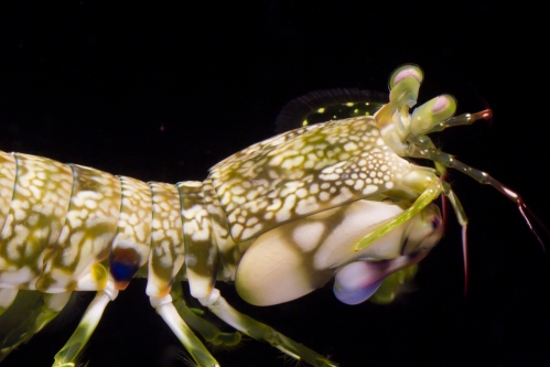
Gonodactylus platysoma (Gonodactyloidea)

Gonodactylus platysoma

I first saw this species of stomatopod in the field this year. They are really beautiful animals, with subtle but vibrant color accents on their dactyls, antennae, eyes, and on the edges of some of their somites (body segments). This individual is about 72 mm in length, and the species seems to be fairly docile (for stomatopods).

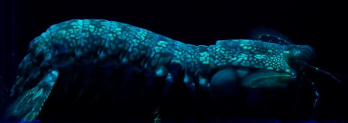
This animal also has very beautiful fluorescent patterns on its body:

G. platysoma; UV-excited fluorescence.

I talked previously about fluorescence in stomatopods here. However, I don’t know if the patterns on G. platysoma are used to amplify any particular signals. These animals live in shallow water and would have less use for fluorescent signal amplification.

Arthropods in pop culture: Attack of the Camel Spiders

Photo: John Sellers

Here is yet another arthropod photo that gets a lot of attention around the internet (and in hysterical mass emails from your aging relatives). During the recent deployments, US servicemen started running into these unsettling arachnids, commonly called camel-spiders, in the Iraqi desert. They subsequently sent home a good deal of photos, rumors, and urban legends about them. To the left is the most popular camel-spider photo that is circulated around the web. Claims about these arachnids include (according to Snopes.com):

  • They are extremely aggressive, viscously charging directly towards, and pursuing soldiers.
  • Running up to 25 mph whilst making a screaming sound.
  • They can grow to be as large as dinner plates.
  • They are able to jump several feet in the air.
  • They are venomous and can anesthetize their prey while they chow down unnoticed.
  • They eat, live in, or lay their eggs in the bellies of camels.

Unsurprisingly, most of this is nonsense fueled by exaggeration, misunderstanding, and wild speculation. Let’s dispel these myths and learn about the interesting reality of Solifugid biology.

Alternately referred to as sun spiders, wind scorpions, and camel spiders; members of Solifugae are neither spiders, nor scorpions, belonging to a distinct evolutionary lineage within the arachnids.

Chelicerate phylogeny adapted from Regier et al., 2010.

Most Solifugids are highly specialized for survival in arid habitats and they are found in deserts around the world, excluding Australia. They are mostly nocturnal to avoid the heat, but some species are diurnal. Shade is crucial to the survival of arid solifugids that are active during the day. The reports of camel spiders charging and pursuing soldiers are likely derived from the animals attempting to take refuge from the sun in their shadows. As far as the shrieking sound that they allegedly make during their charges: Perhaps the sound actually comes from the men, who later try to save face in front of their buddies by attributing the noise to the arachnid. I can relate.

One of the most obvious physical characteristics of solifugids are their massively enlarged chelicerae. These appendages give the impression of tremendously engorged, venom-laden fangs. However, their size is actually a compensation for a lack of venom (There is a single Indian species, Rhagodes nigrocinctus that may possess venom glands, but this has not been well confirmed and there is no known injection mechanism). Each of the chelicerae are composed of two segments forming powerful pincers. These pincers are used to grasp and tare apart their prey; which includes other arthropods, lizards, snakes, and possibly small mammals. Solifugids do not feed on animals larger than themselves, and they do not munch away on humans or camels, unnoticed through the use of anesthetic venom. If these guys take a chomp out of you, you will notice. However, they are not particularly aggressive to people unless harassed or backed into a corner.

The chelicerae of solifugids. From Punzo, 1998; and I. Lindsey

Unlike spiders and scorpions, solifugids superficially appear to possess ten legs. However, the largest, foremost ‘legs’ are actually enlarged, antennae-like, sensory appendages, called pedipalps. The pedipalps are also used for climbing and prey capture.

Galeodes arabs, redrawn by Richard Fox, from Savory, 1977.

Additionally, the first set of true legs are also used as accessory sensory appendages, leaving only the back three sets of legs for locomotion. Solifugids are nonetheless capable of quick bursts of speed (up to 53 cm/sec or 1.2 mph) when attacking prey or darting for cover. However, the claims that they can travel at 25 mph and keep pace with Humvees are obvious exaggerations.

Now, to address the ‘size of dinner plates’ claim about solifugids, which is reinforced by the popular image at the top of the article: Amazingly, for these sorts of meme inducing images, I was actually able to track down the photo’s origin. According to Paula Cushing, Department Chair and Curator of Invertebrate Zoology at the Denver Museum of Nature and Science, the photo was taken by a serviceman and amateur photographer named John Sellers. He photographed the two animals, one violently clamped onto the others abdomen, following a gladiatorial battle between the two solifugids staged by the soldiers (an ugly practice that has gone on at least since British troops were stationed in Egypt during the first World War, and continues today in Youtube videos). Though I was unable to determine an exact species ID, the pictured solifugids are members of the Galeodes genus (perhaps G. granti or G. arabs?). They are about 10 cm in total length, residing at the upper end of the solifugid size spectrum, which ranges from 1 cm to 10 cm in body length. The largest example of a solifugid I could dig up is this Galeodes fumigatus, which appears to be about 11 cm or more in body length. So these animals can reach a menacing size, but nothing close to a dinner plate, or the size suggested by the tricky perspective of the camel-spider meme photo.

Some solifugids can be large and creepy-looking, to be sure, but they are not the deadly, bloodthirsty, lightning-fast, venom-dripping, monsters that popular culture has portrayed them as. If you can redirect fear and revulsion into fascination, and look beyond the unsettling facades of monstrous arthropods, you may find a vibrant wealth of astonishing realities.

Photo by Mundo Poco


Previous posts about arthropods in popular culture:

References:

Thanks to Paula Cushing and Mark Harvey for additional assistance.

  • Punzo, F. 1998. The Biology of Camel Spiders. Kluwer Academic Publishers, Norwell, MA, USA.
  • National Geographic
  • Solpugid.com
  • Snopes
  • Aruchami, M & Rajulu, GS. 1978. An investigation on the poison glands and the nature of the venom of Rhagodes nigrocinctus (Solifugae: Arachnida). Nat. Acad. Sci. Letters (India) 1: 191–192.

Calanus finmarchicus

Check out this cute, red-pigmented, copepod.

Calanus finmarchicus

Awwwwww.

Aphid adornment: Lateral gene transfer from fungi to aphids.

Carotenoids are integral components of animal biochemistry. These organic compounds, characterized by long hydrocarbon chains and loops, are used in photoreception, antioxidation, the immune system, and for ornamental coloration. There are over 800 known carotenoid compounds found in nature. They absorb varying wavelengths of blue and green light, causing tissue containing large quantities of carotenoids to appear green, yellow, orange, or red. This absorptive property is what makes carotenoids crucial for vision and coloration in animals.

However, there is a snag. Animals cannot produce their own carotenoids. Their genomes lack the enzymes necessary to synthesize carotenoids from smaller hydrocarbons, and therefore they must ingest carotenoids from organisms that can. Certain bacteria, archea, plants, and fungi are all capable of producing specific carotenoids. By adding these organisms to their diet, animals can fulfill their carotenoid needs.

One animal that takes advantage of carotenoid coloration is the pea aphid, Acyrthosiphon pisum. There are two primary color morphs of this aphid species, green and red (see below). The green aphid morph has large quantities of greenish-yellow carotenoids: alpha-, beta-, and gamma-carotene. The red aphid has decreased amounts of those carotenoids, and instead has gobs of the red carotenoid, torulene.

Color morphs of the pea aphid, A. pisum, and the carotenoids predominately responsible for the color variation. Adapted from Moran and Jarvik, 2010.

This polymorphism is of great ecological significance for the aphid. Parasitoid wasps prefer to lay their eggs in green aphids, and carnivorous ladybugs prefer to eat red aphids. If there is a spike in wasp parasitism, the red morphs become more predominant. If there is a spike in ladybug predation, then the green morphs become more common. By maintaining the genetic diversity of both color morphs in an aphid population, that population can guard against being wiped out by a temporal increase in parasites or predators.

However, this polymorphic color variation seen in aphids presents an interesting question. Where are the aphids getting their carotenoids? The plant phloem sap that aphids suck out of leaves is low in carotenoids, and the carotenoids produced by aphid host plants do not match those found in the aphids. In addition, the endosymbiotic bacteria within the aphids cannot be the source of the carotenoids, as there are no carotenoid biosynthesis genes in their genomes.

However, researchers were surprised to discover that the aphid’s red-green color polymorphism is inherited in a classic Mendelian autosomal manner (remember your Punnett squares, kids?), with the red allele dominant to the green allele. This indicates that the genes responsible for carotenoid-based colorations in these aphids are located within their own genomes! A search of the newly published pea aphid genome revealed the presence of several carotenoid synthases, cyclases, and desaturases. This suite of carotenoid biosynthesis genes is capable of producing the colored carotenoids necessary for the red and green aphid color morphs. A mutation in one allele of these genes prevents the production of the red carotenoid, torulene, resulting in the green aphid color morph.

These are the first carotenoid biosynthesis genes found in an animal. Indeed, no other known animal genome, including several other insect genomes, contain homologues to these genes: so where did they come from?

Carotenoid synthesis genes from various organisms. Adapted from Moran and Jarvik, 2010.

A gene tree containing the aphid carotenoid genes, as well as similar genes from bacteria, plants, and fungi can bee seen to the left. The aphid genes (blue) are completely nested within fungal carotenoid biosynthesis genes (brown). Therefore, at some point in aphid evolution, the genes for carotenoid biosynthesis were transfered from a fungi directly into the aphids genome. It is likely that a similar event occurred from bacteria (black) to plants (green) as well. The possible mechanisms for such gene transfers, especially between two eukaryotes, are poorly understood at this point.

Earlier, I wrote about a similar case of lateral gene transfer from algae to a sea slug. That was the first known transfer of genes between multicellular organisms. This new example in aphids lends support to the notion that these lateral gene transfers are more common in eukaryotes that once though. It may turn out, as genome sequencing increases at exponential rates, that the eukaryotic tree of life actually resembles (to some extent) the interconnected gene web seen in bacteria.

References:

  • Moran, N., & Jarvik, T. (2010). Lateral Transfer of Genes from Fungi Underlies Carotenoid Production in Aphids Science, 328 (5978), 624-627 DOI: 10.1126/science.1187113

Hat tip to Microecos for tweeting this.

I want my own enormous robotic ant

Unleash the awesome!

A robotic engineer has developed a beautifully designed hexapod robot based on the biomechanics of ants. He calls his remote controlled creation A-pod, and cites the photography of Alexander Wild (of Myrmecos) as an influence on his design.

A-pod is capable of a wide range of motions and body contortions. In addition to walking around, A-pod can grasp and carry objects in its metallic mandibles. The motions are incredibly fluid and I can’t imagine the amount of work that had to go into programming the synchronous movements of all six legs. You can watch videos of A-pod in action here and here, and learn more about the construction of the robo-beastie here.

At this point, A-pod just needs to be programmed to find Sarah Connor, and then it can assist in the inevitable robot uprising.


I have moved.
Arthropoda can now be found here.

Michael Bok is a graduate student studying the visual system of mantis shrimp.

Flickr Photos